 +5 more
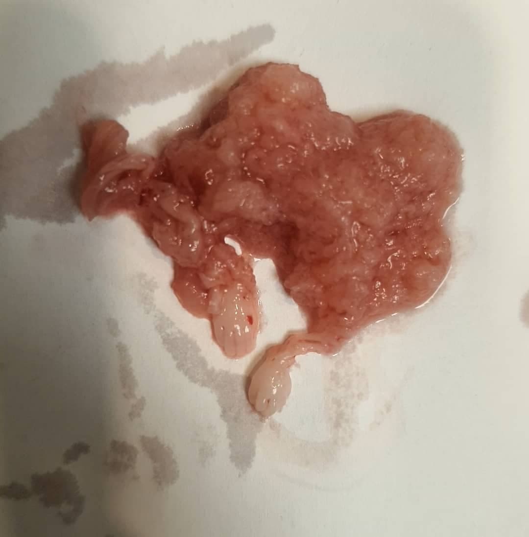
A 50 y/o female with a cecal polyp (0.7cm). Dx: Sessile serrated lesion (also known as sessile …
professionaly/ofemale
August 18, 2024
+5 moreA 50 y/o female with a cecal polyp (0.7cm). Dx: Sessile serrated lesion (also known as sessile …
 +8 more
A 60 y/o male with a scrotal mass. Dx: Scrotal Calcinosis
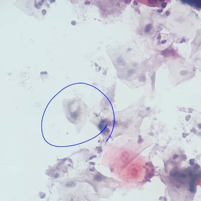
 +4 more
Routine pap smear Presence of squamous cells with enlarged hyperchromatic nuclei and irregular …
 +5 more
Well-differentiated SCC of tongue, keratinizing With entrapment of skeletal muscle bundles …
 +9 more
A 46 y/o woman with a sole lesion. A well-defined mass with epithelial lobules composed of two …
 +2 more
Early secretory endometrium, resembling piano keys
 +1 more
Adenomyomatous hyperplasia of gallbladder
 +5 more
Frightening LSIL in pap smear 😱
 +7 more
Fibrin clumps in shedding endometrium
 +2 more
This is how the gestational endometrium looks like! 🤗
 +2 more
Diverticulum of appendix, not an everyday case !
 +4 more
Resected as "Meckel diverticulum" On gross examination, an ill-defined polypoid elevated area on …
 +9 more
A 33 y/o female with a polypoid pendunculated vulvar mass A lesion with vascular and myxoid …
 +9 more
An 11 y/o male with a lower lip lesion A cyst like structure lined by abundant foamy histiocytes …
 +9 more
A 35 y/o female, asymptomatic with a liquid based pap smear for routine screening Very slight …
 +4 more
A 64 y/o female with a lesion on face Skin tissue with papillomatosis, acanthosis and …
 +5 more
An 85 y/o male with an old skin lesion on leg A quite symmetrical skin lesion with …
 +8 more
A 46 y/o female with complaint of AUB, sonographic exam showed an endocervical polyp with …
 +1 more
Alternaria in pap smear! Usually due to staining contamination
 +7 more
And here it is.. bilateral pilomatrixoma! An 8 y/o boy with bilateral soft tissue lesions at the …
 +7 more
IUFD in a 27 y/o female (GA: 32w) Infiltration of neutrophils in chorion, early chorionitis/acute …
 +3 more
Cutie cholesterolosis 😍
 +2 more
A 37 y/o female with thigh skin lesion. Dx: Molluscum contagiosum and intradermal melanocytic …
 +9 more
Terrifying poorly differentiated gastric adenocarcinoma with signet ring features.. Marked …
 +5 more
A 21 y/o female with vaginal discharge and pruritis, no history of recent lactation or pregnancy, …
 +3 more
Pigmented lesion on face and suspicion of malignancy... Dx: Pigmented seborrheic keratosis
 +3 more
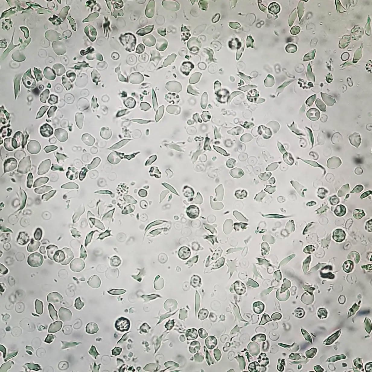
Eye catching ammonium biurate crystals in urine analysis @faeze_m94
 +2 more
Here they are..weird shapes of atypical mitosis
 +5 more
Hard to believe that the slides are from peripheral blood and not bone marrow! A 35 y/o male with …
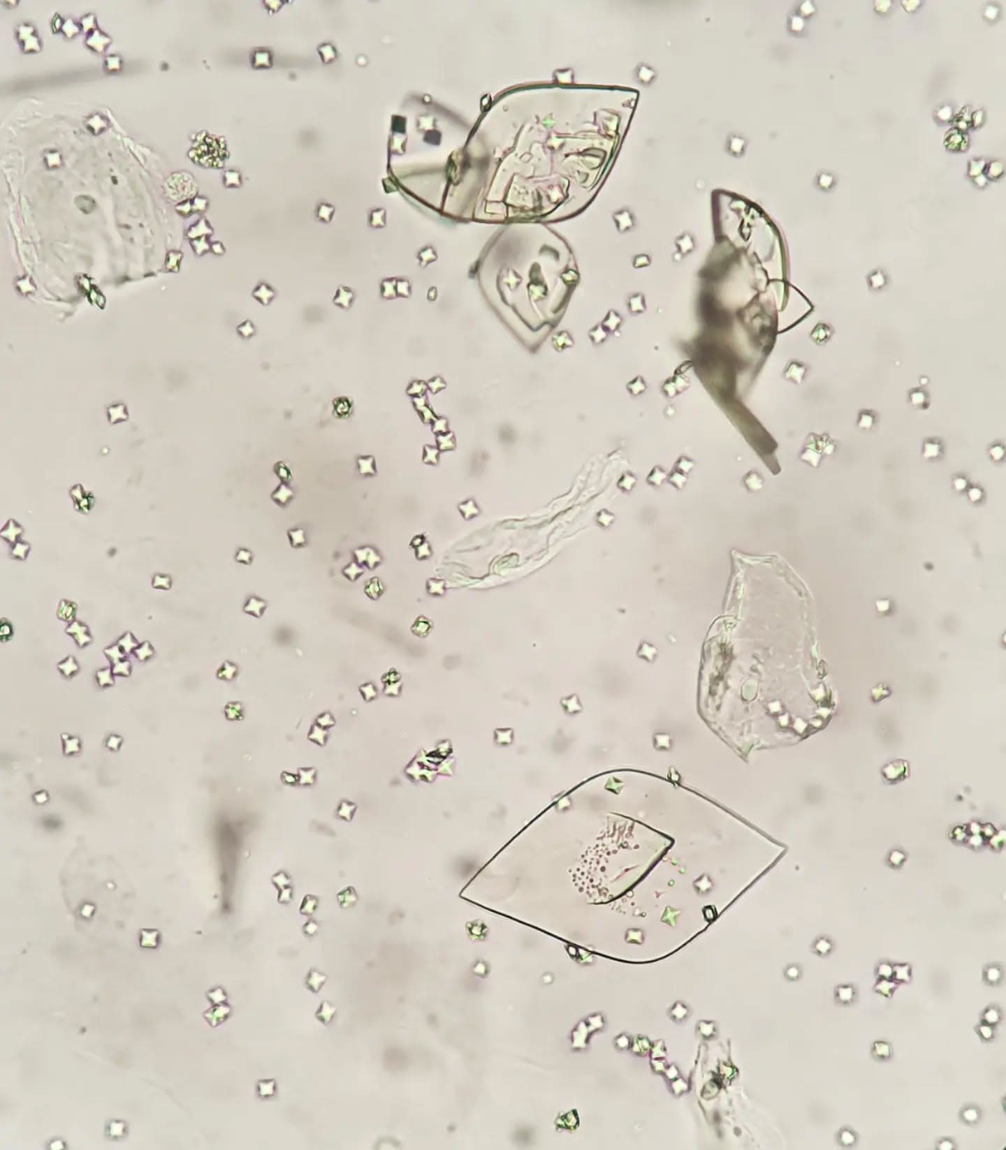
Urine analysis, microscopic exam, many amazing calcium oxalate and some uric acid crystals …
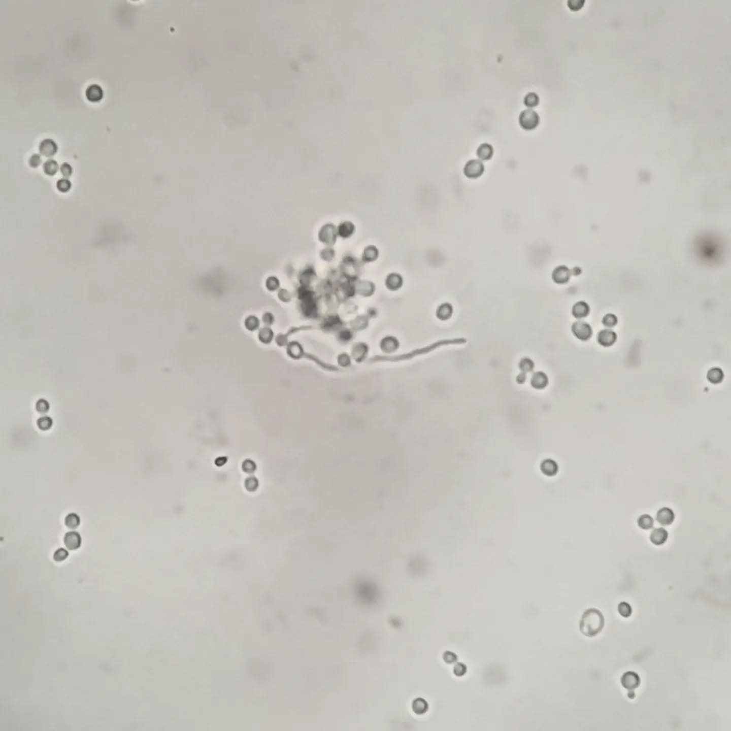
Amazing germ tube test performed on urine specimen indicative of candida albicans
 +2 more
A case of beta thalassemia major who has undergone splenectomy. On peripheral blood smear howell …
 +4 more
A case of sickle cell anemia with Hb: 7.1, MCV: 85, MCH: 27, MCHC: 31, WBC: 11'400, plt: 233'000. …
Liver mass On cutting, many daughter cysts came out of the so-called mass, making the diagnosis …
 +1 more
How acute cholecystitis may look like in gross exam with diffusely hemorrhagic mucosa and a large …
 +9 more
Lung adenocarcinoma with involved lymph node
 +8 more
History of uterine leiomyosarcoma and metastasis to lung, again with multiple lung …
 +2 more
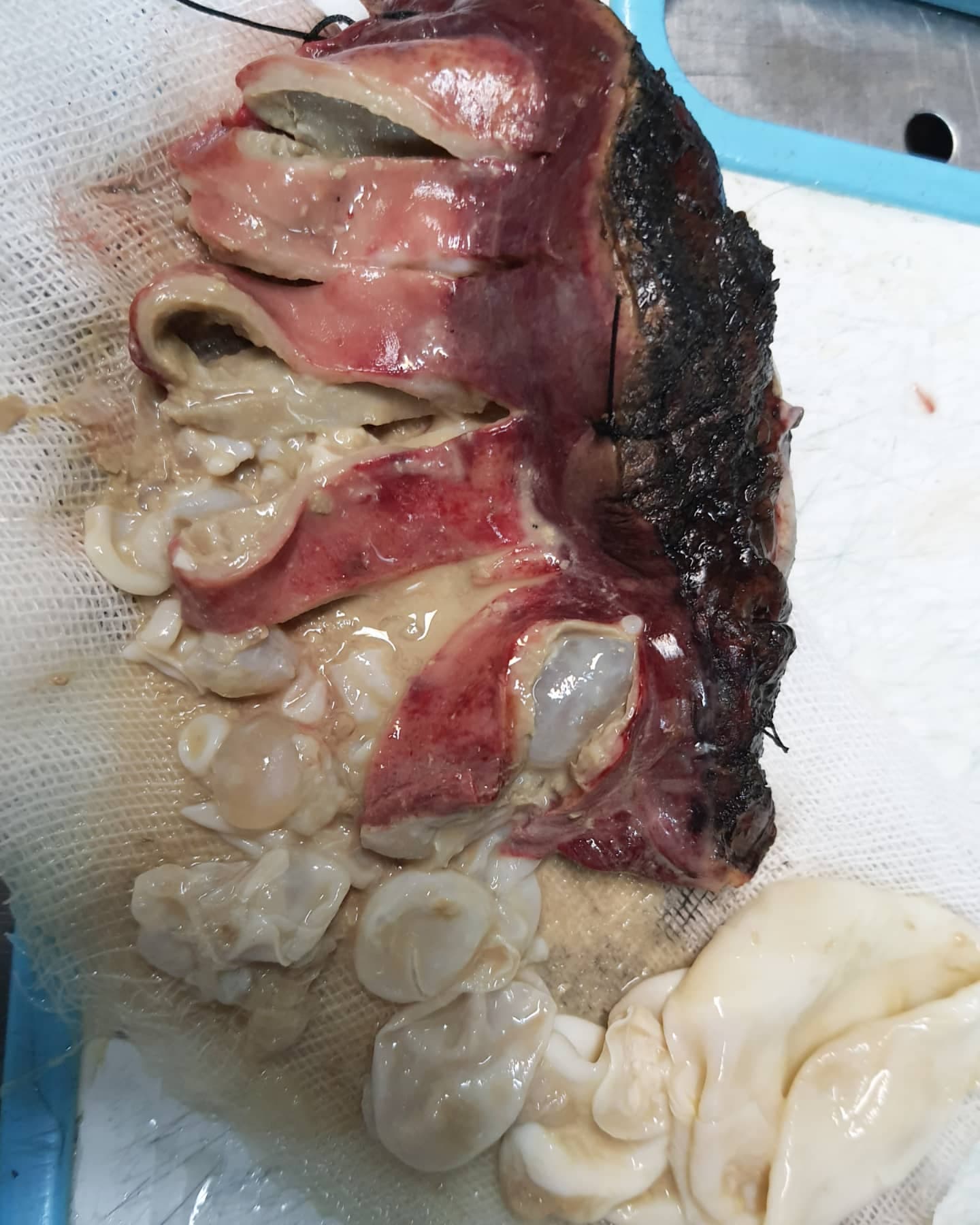
How a lung with bronchiectatic changes looks like in gross exam..distended peripheral bronchi …
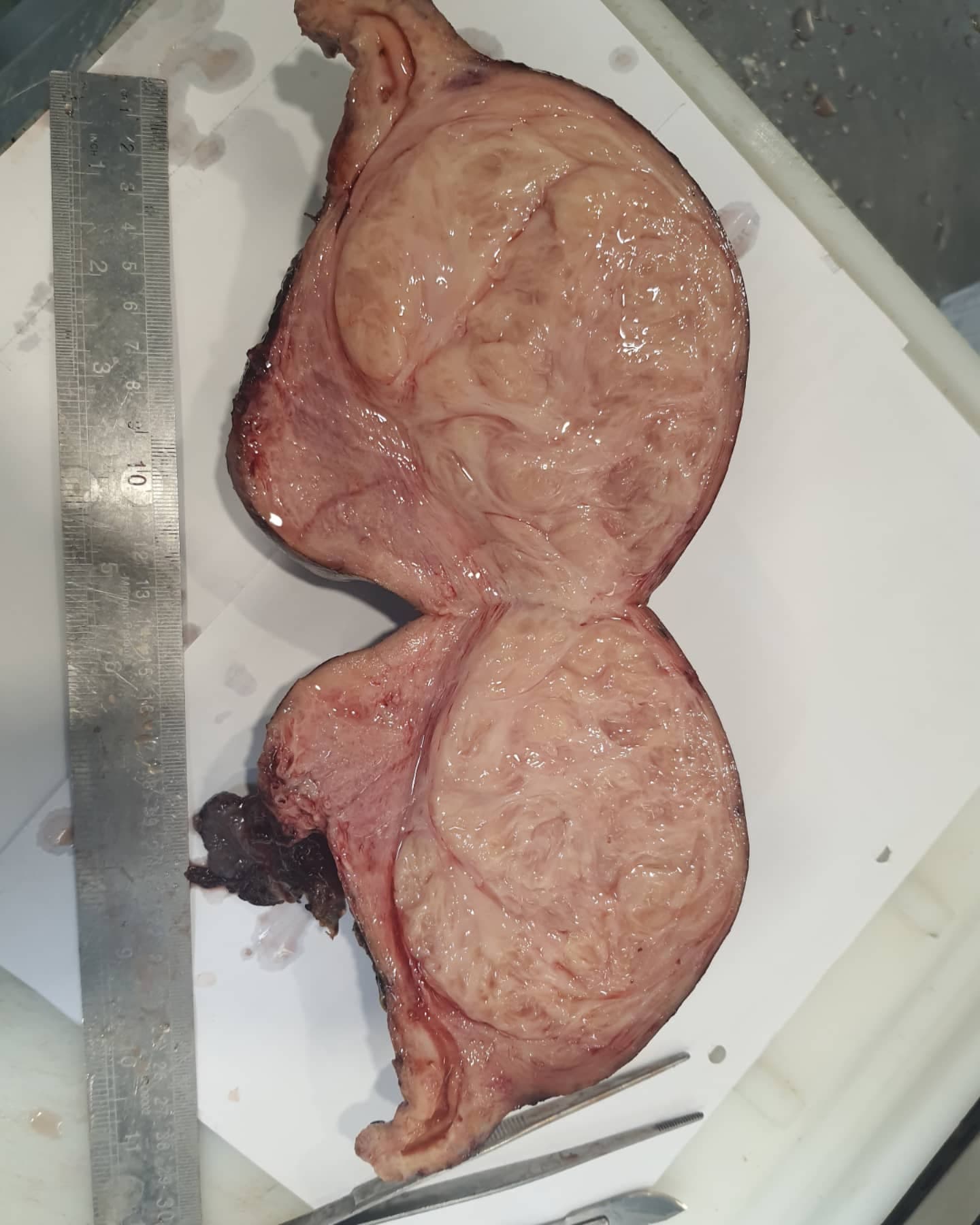
Very large intramural myoma (M: 12cm in greatest dimension) with nice whorled pattern in cut section
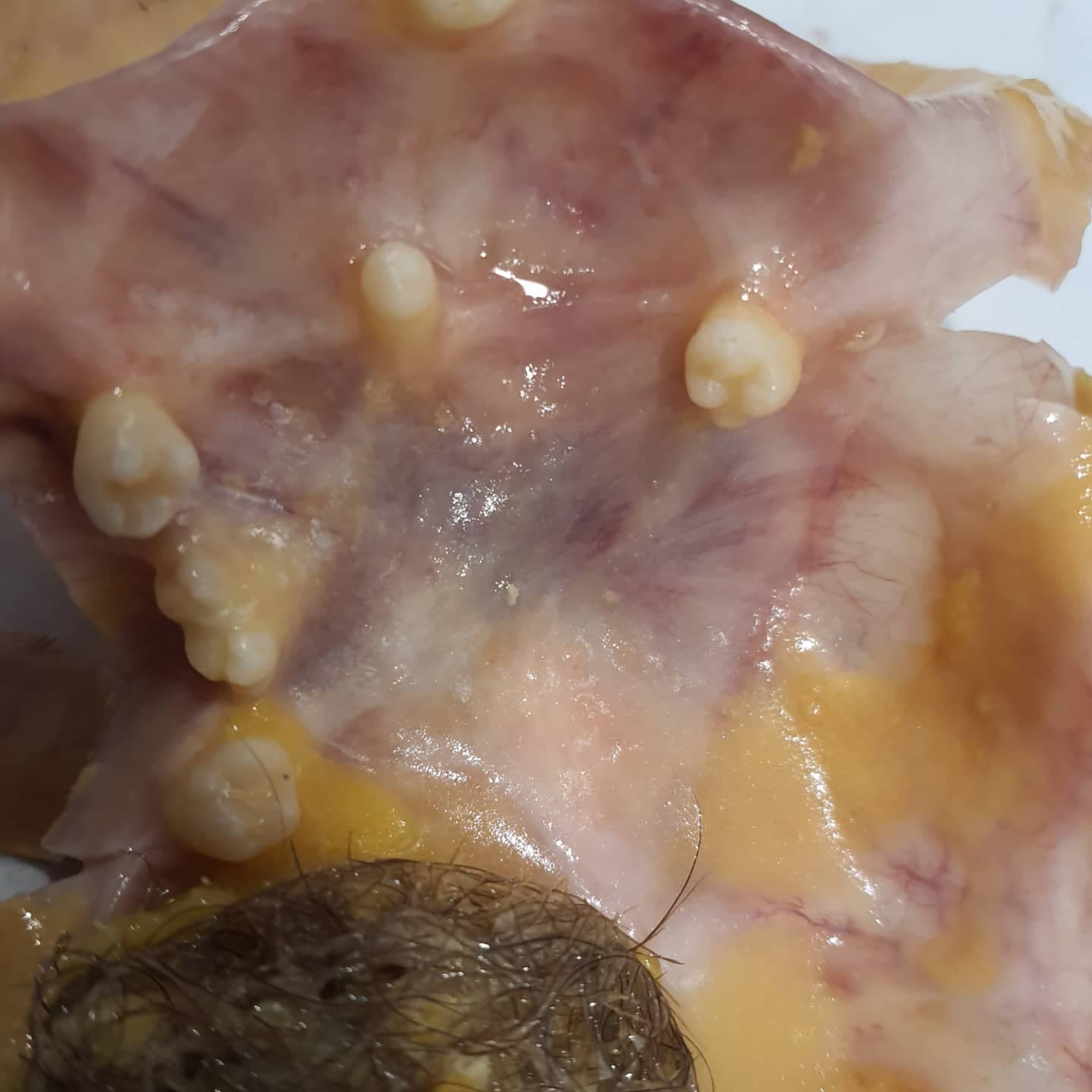
How many well formed teeth have you ever seen in a mature cystic teratoma? 😁😄
 +1 more
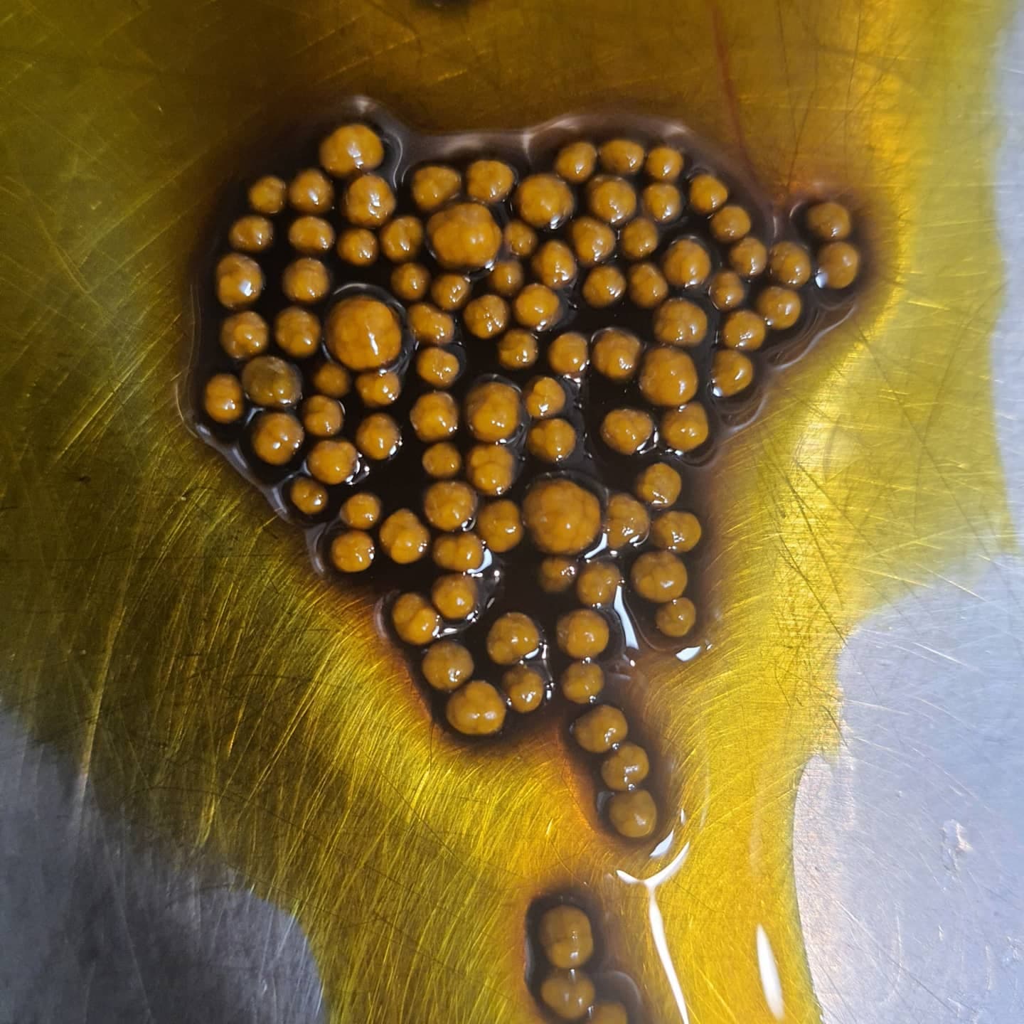
Nice monomorphous gallbladder stones with impeccable resemblance to a bunch of pearls
These very tiny hands/feet-to-be.. P.S. D&C specimen